
Multiscale Imaging
Visualizing ultrasound-mediated processes from microbubble dynamics to cellular and vascular responses.
Our laboratory develops and applies optical imaging approaches to investigate ultrasound-mediated physical and biological processes across spatial and temporal scales.
High-frame-rate microscopy enables us to resolve rapid microbubble dynamics and their interactions with cells and biological interfaces, linking acoustic forces to microscale mechanical effects.
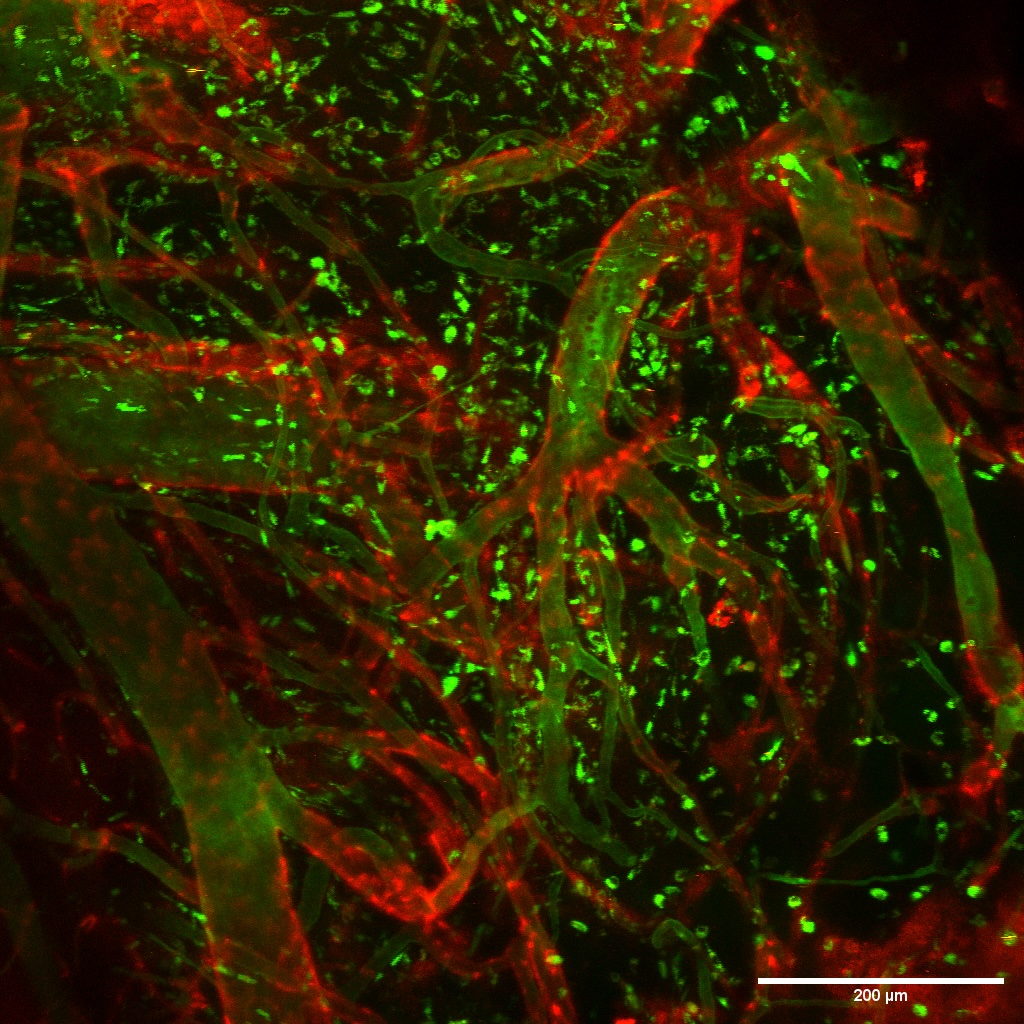
Multiphoton intravital microscopy extends these observations in vivo, allowing real-time visualization of vascular dynamics and immune-cell trafficking in the mouse brain. By fluorescently labeling the vasculature and immune cells, we can track individual cells and resolve key steps in the extravasation cascade.
Ultrasound Imaging – Beyond microscopy, we develop and apply advanced ultrasound imaging methods to noninvasively monitor microbubble activity, tissue perfusion, and guide therapeutic intervention in real time. These approaches provide quantitative imaging biomarkers that guide therapeutic ultrasound procedures and connect microscale mechanisms with clinically relevant imaging endpoints.
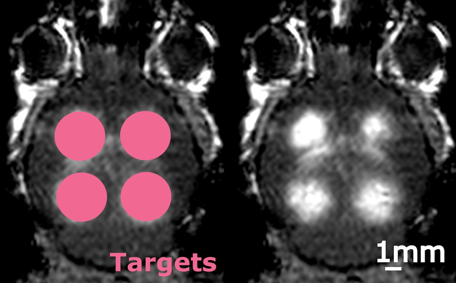
Magnetic Resonance Imaging (MRI) – MRI plays a central role in validating and translating our therapeutic ultrasound approaches, enabling longitudinal assessment of blood-brain barrier opening, drug delivery, and treatment response. By combining MRI with ultrasound and optical imaging, we establish multiscale imaging frameworks that bridge cellular mechanisms with whole-brain and clinically relevant outcomes.
Together, these complementary approaches connect microbubble dynamics with cellular and vascular responses, providing mechanistic insight that guides the design of focused ultrasound strategies for therapeutic delivery and immune-cell modulation.