Quantitative Immune-Cell Imaging
Understanding and exploiting intracellular microbubble dynamics to image immune-cell trafficking in vivo.
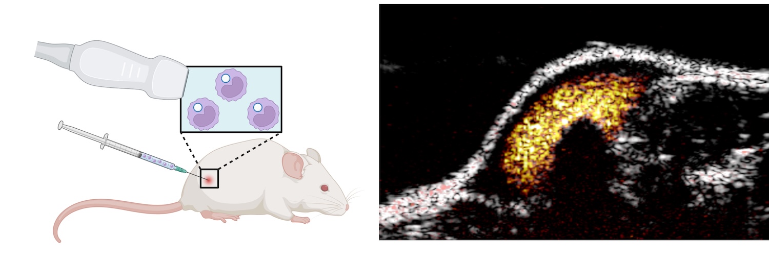
Monitoring immune-cell trafficking remains challenging because it requires high sensitivity and specificity while preserving spatial resolution and ability to image at different depth. We discovered that although cellular compartments damp microbubble (MB) oscillations, internalized MBs retain their nonlinear behavior under low mechanical-index sonication, enabling highly specific nonlinear ultrasound imaging against predominantly linear background tissue. We established non-destructive methods for labeling macrophages with MBs without altering cell viability, migration, phenotype, or cytokine secretion. This approach achieves single-cell sensitivity in vitro and enables longitudinal monitoring of macrophage accumulation in solid tumors in vivo following intravenous administration (Nature Communications, 2025), providing a platform for quantitative imaging of immune-cell trafficking.
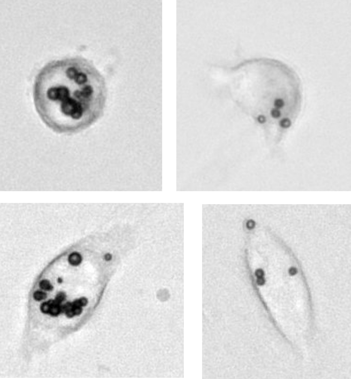
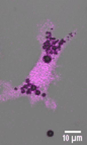
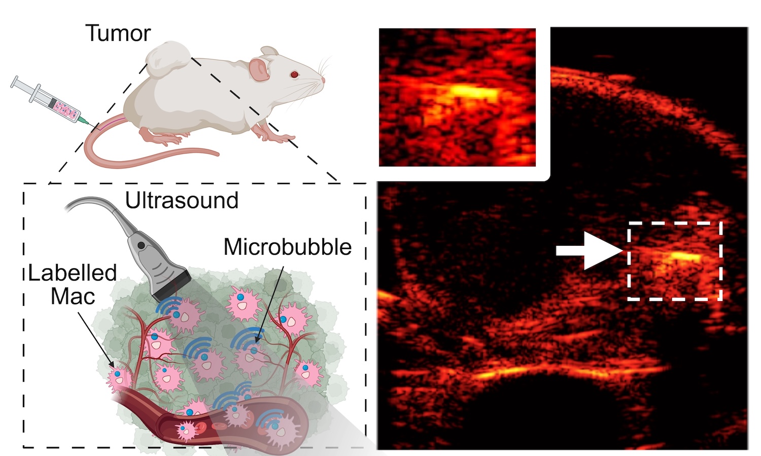
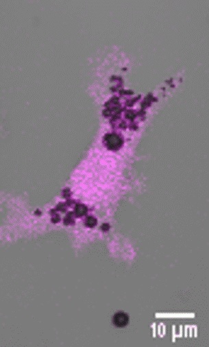
Building on our macrophage imaging work, we have extended our labeling approach to human macrophages and dendritic cells, broadening its translational relevance. In parallel, we have been investigating what happens to microbubbles once internalized by immune cells, which is a key challenge for longitudinal cell tracking. Using live cell microscopy, multigas diffusion modeling, and ultrasound imaging, we found that intracellular MB dissolution is driven primarily by gas efflux rather than enzymatic degradation (Theranostics, 2026). Saturating the surrounding medium with perfluoro-n-pentane (PFP) to compensate core gas diffusion significantly extended intracellular MB persistence both in vitro and in vivo. This work establishes a mechanistic framework for studying the lifetime of internalized microbubbles and advancing longitudinal ultrasound immune cell tracking.